Osteology:

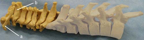
The temporal bone, zygomatic arches, occipital bone, mandible, cervical vertebrae, scapula clavicles, manubrium, and hyoid bone form the skeleton of the neck.

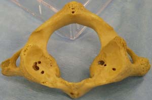
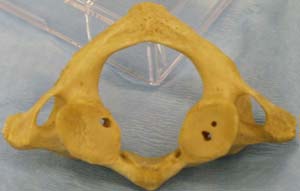
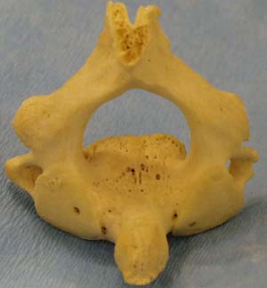
Cervical Vertebrae: C1-C7


    1. small and longer from side to side than anteroposteriorly
    2. superior surface is concave forming the uncinate processes laterally
    3. inferior surface is convex
    4. can be used as a landmark for hyoid (C3), thyroid cartilage and bifurcation of the common carotid artery (C4), cricoid cartilage (C6)

Hyoid Bone

Surface Anatomy and Other Landmarks

  1. Laryngeal prominence (Adam's apple) in the midline formed by the thyroid cartilage at approximately C4.
  2. Inferiorly the ring of the cricoid cartilage may be palpated at C6.
  3. The tips of the transverse processes of C1 are more prominent than those of other cervical vertebrae and can be palpated in the parotid space.
  4. The hyoid bone: Its body is at the level of C3. It has lesser and greater horns bilaterally.
  5. The thyroid cartilage lies at the levels of C4 and C5. The laryngeal prominence is Adam's apple. The thyroid cartilage is composed of 2 lateral laminae with superior and inferior horns. The inferior horns articulate with the cricoid cartilage.
  6. The cricoid cartilage at C6. The upper end of the trachea is palpable in the midline from the cricoid cartilage to the superior border of the manubrium.
  7. The thyrohyoid membrane is pierced by the internal laryngeal nerve and vessels.
  8. The cricothyroid membrane may be used for a high tracheostomy.

*The preferred site of tracheostomy is at tracheal cartilages 2-4 (below cricoid cartilage and isthmus of the thyroid gland).

BACK